

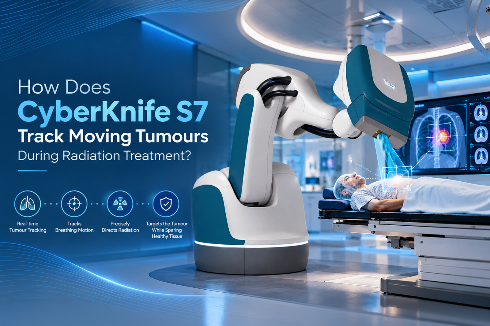
“Will radiation still reach my tumour when I breathe?” This is an understandable question, especially for patients with lung, liver or pancreatic tumours.
CyberKnife S7 addresses this challenge by combining X-ray imaging, external breathing sensors, motion-modelling software and a robotic treatment arm. Its Synchrony technology estimates where a moving tumour is between imaging checks and adjusts radiation delivery accordingly. This helps the treatment beam follow the target while the patient breathes naturally.link.
At Valentis Cancer Hospital in Meerut, CyberKnife S7 forms part of the hospital’s advanced radiation treatment services. Understanding its tracking process can help patients approach treatment with greater clarity.
Why Do Tumours Move During Radiation Treatment?
When you breathe, your diaphragm moves and your chest expands. These movements can shift organs inside the chest and abdomen, including the lungs, liver and pancreas. A tumour within these organs may therefore change position throughout the breathing cycle.
Radiation treatment for moving tumors must account for this movement. Without appropriate motion management, a plan may need a larger treatment margin around the tumour to accommodate its changing position.
CyberKnife’s respiratory tracking aims to follow this motion, helping clinicians target the tumour while limiting unnecessary radiation exposure to nearby healthy tissue. Safety margins remain necessary; tracking does not eliminate uncertainty entirely.
How CyberKnife S7 Tracks a Moving Tumour
The process involves several connected technologies rather than a single camera watching the tumour continuously.
1. X-Ray Images Locate the Internal Target
During treatment setup, paired X-ray images help establish the target’s position. Depending on the treatment approach, the system may identify the tumour directly or locate implanted markers called fiducials that serve as reference points.
Some suitable lung tumours can be tracked without implanted markers. Whether marker-free tracking is possible depends on how clearly the target appears on the imaging system.
2. External Sensors Monitor Breathing
Small light-emitting markers placed on the patient’s chest or abdomen allow an optical camera system to monitor external breathing movement.
These markers do not directly detect cancer. Instead, they provide a continuous signal showing how the body’s surface moves during breathing. The system links this signal with internal target positions obtained from X-ray images.
3. Synchrony Builds a Motion Model
The software creates a mathematical relationship, called a correlation model, between external breathing movement and internal tumour movement.
For example, it learns where the target tends to be as the patient breathes in and out. This relationship allows the system to estimate the tumour’s position between X-ray checks.
Importantly, the model can account for differences between inhalation and exhalation rather than assuming that internal movement always follows a simple, identical path.
4. The Robotic Arm Adjusts Beam Direction
Using the estimated target position, the robotic arm adjusts radiation delivery to follow the moving target.
Think of it as carefully aiming at a moving object rather than repeatedly treating a fixed point and hoping the object returns there. However, this analogy simplifies a tightly controlled medical process: tracking accuracy depends on imaging, modelling, planning and clinical checks working together.
5. Fresh Images Check the Model
Breathing patterns can change during a session. The system therefore acquires additional X-ray images periodically to check and update the relationship between external movement and internal target position.
This distinction matters: continuous external monitoring is combined with intermittent internal imaging, not continuous X-ray filming of the tumour.
What Are the Benefits for Patients?
For appropriately selected patients, motion-synchronised robotic radiation treatment offers several potential advantages:
- Natural breathing during respiratory-tracked delivery, often without repeated breath-holding.
- Beam adjustments that account for tumour movement.
- Focused treatment designed to reduce exposure to surrounding tissue.
- A non-incisional treatment approach.
The CyberKnife S7 manufacturer’s brochure describes Synchrony respiratory modelling as keeping the beam aligned with the target during normal breathing, including irregular breathing patterns. Nevertheless, individual suitability and treatment accuracy require professional assessment.
Does Every Patient Need This Tracking Method?
No. Tracking is selected according to the tumour’s location, visibility and movement. A respiratory-motion model may be useful for a liver tumour but is not automatically needed for every brain or spine treatment.
Patients exploring cyberknife treatment in India should ask whether their plan requires fiducial placement. Although radiation delivery is non-incisional, placing fiducials is a separate procedure when needed. Marker-free tracking is suitable only in selected circumstances.
Choosing Care at Valentis Cancer Hospital
Technology works best when supported by careful planning and an experienced clinical team. Patients choosing a cancer hospital in Uttar Pradesh should ask how motion will be assessed, which tracking method is proposed and how treatment accuracy will be checked.
Valentis Cancer Hospital offers CyberKnife S7 in Meerut. A specialist consultation can clarify whether this technology suits your diagnosis and how its motion-tracking capabilities would apply to your treatment plan.
